How Talus Misalignment Can Lead to Bunions: A Manual Therapist’s Guide
For attendees of the Feet Mechanics Workshop
By Riccardo Galeotti | The Body Lab | 8 May 2025
Why does talus misalignment matter in bunion formation?

Bunions (hallux valgus) are more than a cosmetic deformity—they’re a biomechanical consequence of whole-foot dysfunction. While genetics, tight footwear, and joint hypermobility are often blamed, a deeper structural issue—talus misalignment—frequently plays a central role.
The talus serves as the keystone of the ankle-foot complex, transferring load between the leg and the foot, guiding pronation/supination transitions, and influencing medial arch function. If it becomes misaligned due to injury, habitual gait compensations, or poor foot control, the body’s centre of mass shifts medially, destabilising the first ray. This sets up the foot for progressive structural failure, including:
First metatarsal drifting medially
Hallux abducting laterally
Increased strain on the medial capsule and sesamoids
As discussed in the Feet Mechanics Workshop and your bunion webinar, this shift begins long before visual signs of bunion appear. You can also watch how to assess the Talus function here
“An unstable or misaligned talus results in altered medial column loading, contributing significantly to hallux valgus formation” (McPoil et al., 2009¹).
How does talus misalignment create bunions biomechanically?
Foot mechanics are a chain reaction. Misalignment of the talus causes:
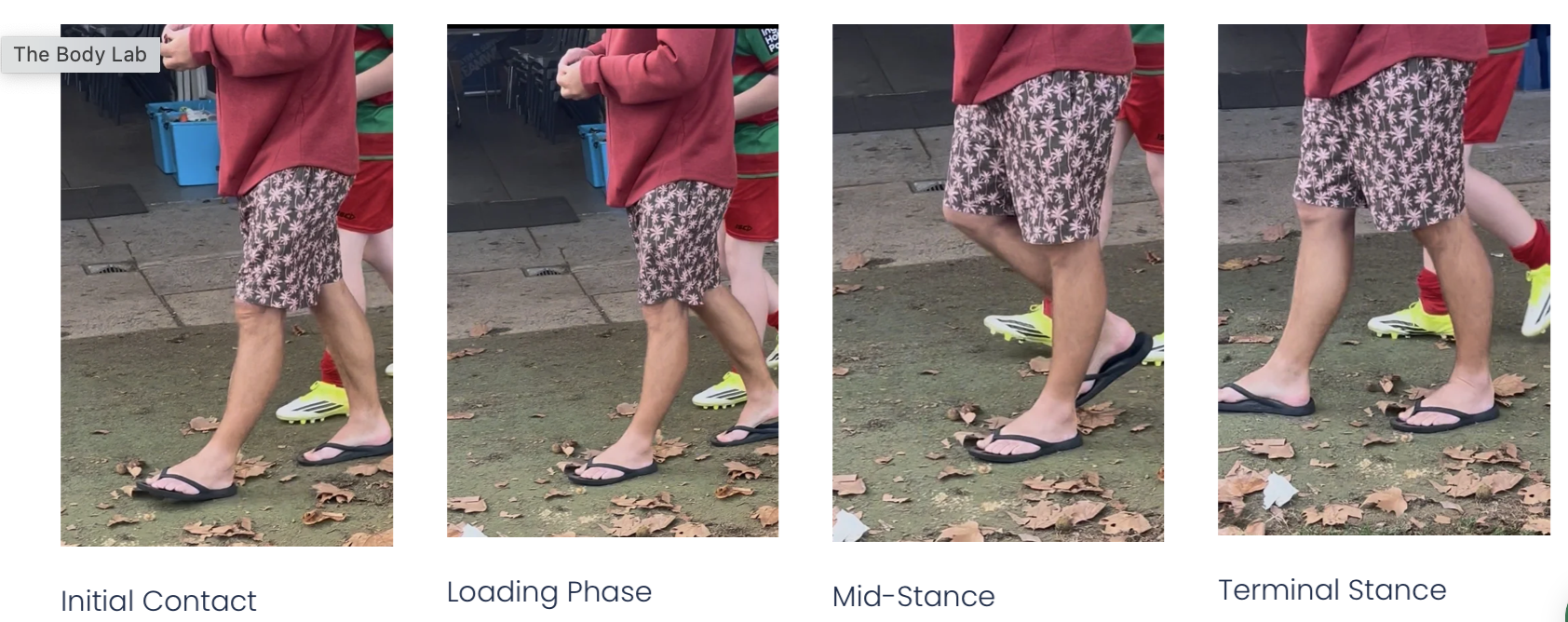
Prolonged pronation: The foot remains unlocked for too long during midstance.
Delayed supination: Without re-supination, the medial arch cannot lift or load effectively for toe-off.
First ray instability: The metatarsal lacks a stable base, causing the big toe to deviate laterally.
Abductor hallucis inhibition: This intrinsic muscle can’t counteract valgus force due to positional disadvantage.
Second toe collapse: Often seen as forefoot instability spreads, shifting more weight medially.
This is reinforced by footwear with narrow toe boxes, which restrict toe splay, dorsiflexion, and natural load sharing between digits. MRI studies confirm that talar positioning influences forefoot abduction torque and sesamoid tracking (Lee et al., 2013³; Kim et al., 2015⁷).
“The subtalar joint’s prolonged eversion and delayed heel rise increase stress on the first ray” (Smith & Katchis, 2013⁶).
When should you suspect talus misalignment in clinical practice?
Red flags for talus dysfunction include:
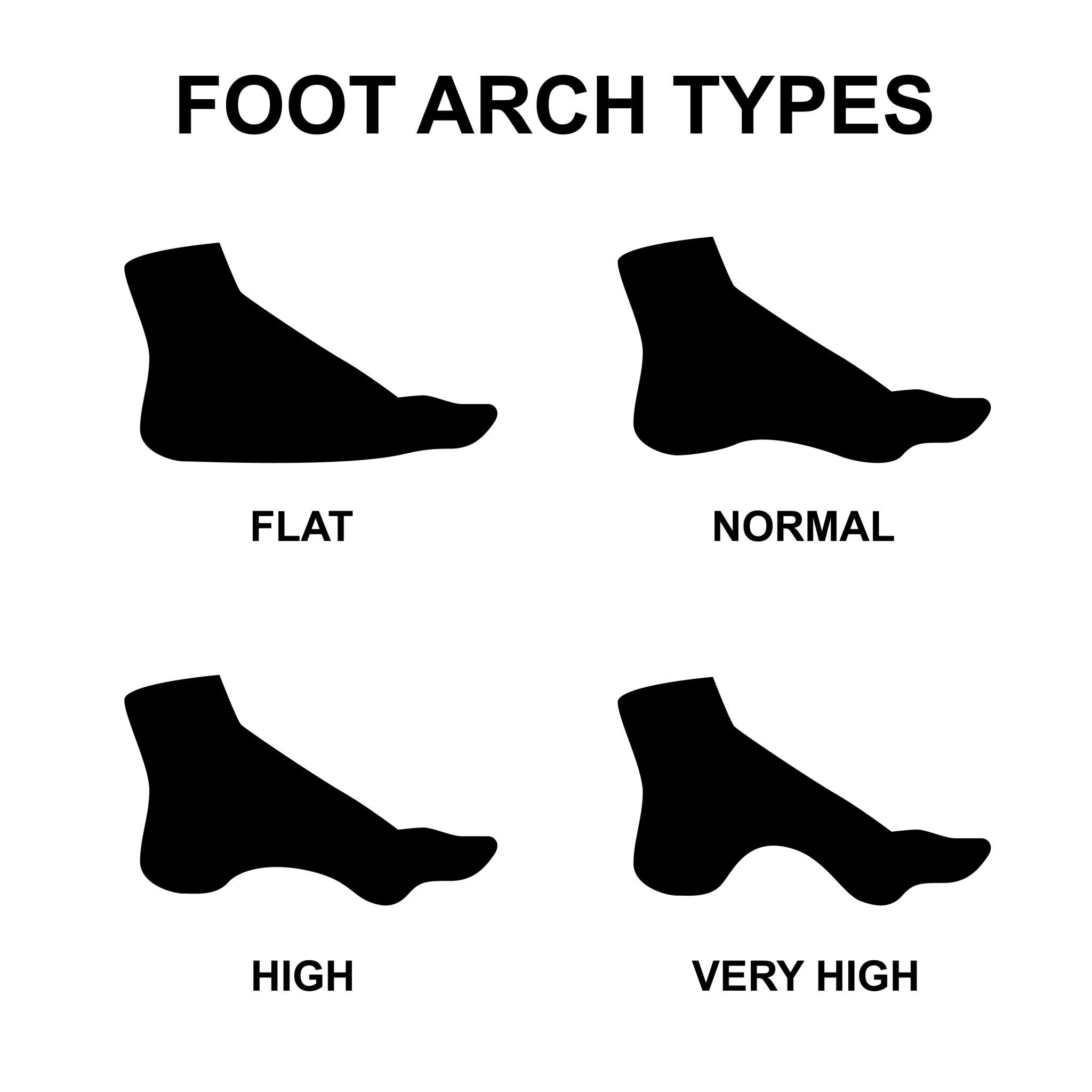
Flat feet or collapsed arches that don’t reappear during supination
Limited ankle dorsiflexion, particularly with eversion
Early heel rise or excessive medial forefoot loading in gait
Callus under 1st or 2nd metatarsals, indicating overload
Sesamoid pain or tenderness at the medial hallux joint
History of ankle sprains, hypermobility, or altered gait
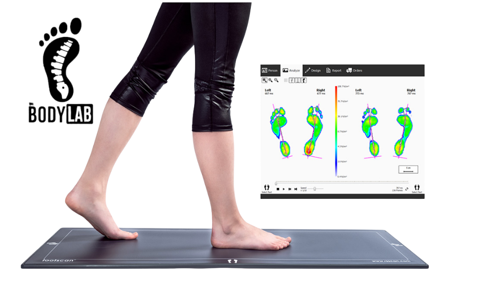
Functional assessments (such as foot tripod tests, single-leg stance, or heel raise assessments) often show the first ray fails to anchor properly or collapses under load. In gait, watch for insufficient toe dorsiflexion during push-off, or a first toe that rotates outward instead of pushing straight ahead.
These signs suggest the talus is failing to guide the subtalar and midfoot joints efficiently, leading to bunion-prone mechanics.
What does this mean for treatment—and what should we do about it?
Treating bunions without addressing talus mechanics is incomplete. Toe spacers, surgery, and orthotics may address symptoms, but long-term success depends on restoring proximal control and joint timing.
Manual therapists should:
Mobilise the talus posteriorly and medially to restore joint glide
Address midfoot mobility (navicular, cuboid, cuneiforms) to re-establish foot tripod
Use functional drills to train pronation-supination sequencing during gait
Activate intrinsic foot muscles (e.g. abductor hallucis) and retrain toe splay
Implement hip and pelvic stability exercises to prevent compensation from above
For clients, understanding that bunions develop over time and can be reversed—or at least slowed—without invasive measures is empowering.
“Mobilisation of the talus and midfoot improves rearfoot alignment and first ray stability” (Vicenzino et al., 2008⁵).
Learn this and more in the Feet Mechanics Workshop
This topic is just the beginning. The Feet Mechanics Workshop covers how the foot moves in all three planes, how dysfunction propagates through the body, and how to use movement, manual therapy, and education to get long-lasting results.
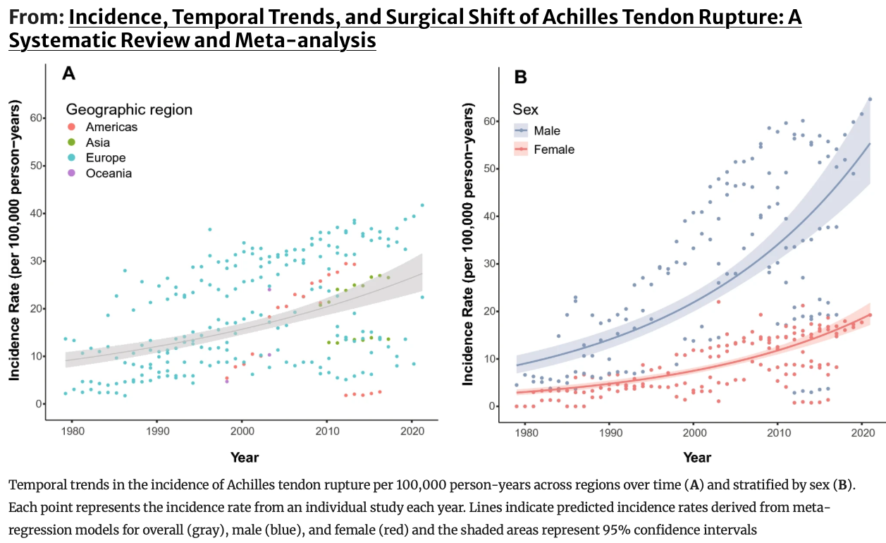
ReferencesMcPoil TG, Cornwall MW. Relationship between foot function and the development of hallux valgus. J Foot Ankle Res. 2009;2(1):11.Dakin SG, Newton J, Martinez FO, et al. Chronic inflammation is a feature of Achilles tendinopathy and rupture. Br J Sports Med. 2018;52(6):359–67.Lee JH, Lee HS, Kim JS, et al. Relationship between first ray hypermobility and the severity of hallux valgus deformity. J Orthop Surg (Hong Kong). 2013;21(2):199–202.Nigg BM, Benno M. The role of joint motion in foot function and pathology. Clin Biomech. 2011;26(5):452–460.Vicenzino B, Branjerdporn M, Teys P, Jordan K. Initial changes in posterior talar glide and pain after mobilization with movement in patients with ankle sprain. Man Ther. 2008;13(5):492–500.Smith RW, Katchis SD. Foot biomechanics and hallux valgus. Orthop Clin North Am. 2013;44(4):405–14.Kim Y, Kim JS, Young KW, Naraghi AM. Radiographic measurements of the first metatarsophalangeal joint in hallux valgus. Foot Ankle Int. 2015;36(3):366–372.