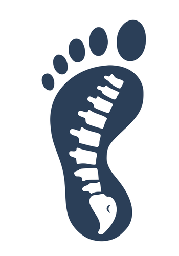
The Foot’s Secret Superpower: Understanding the Windlass Mechanism
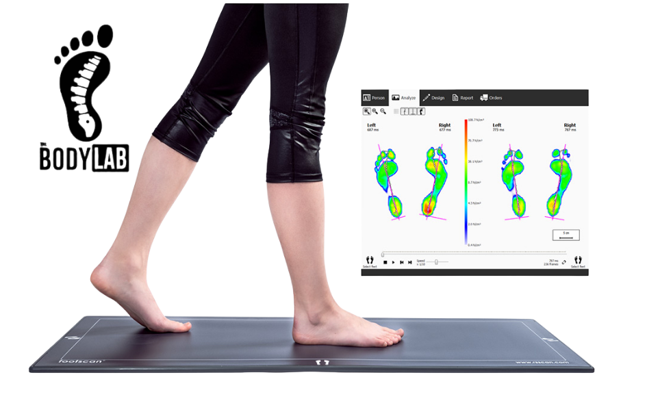
The Body Lab Canberra
(Keywords: foot pain Canberra, plantar fascia, windlass mechanism, foot mobility exercises, biomechanics of gait)
Introduction: The Sail Hoist in Your Sole
Picture this: a sailor cranks a pulley to hoist a heavy sail. With every turn of the handle, the rope tightens, the load lifts, and the system suddenly feels effortless. That pulley is called a windlass—a device that turns tension into mechanical advantage.
Now, here’s the twist:
There’s a windlass inside your foot.
It’s called the Windlass Mechanism, and it’s powered by a humble strip of tissue known as the plantar fascia—that taut, fibrous band running from your heel to your toes. This built-in pulley system is what transforms your foot from a soft, shock-absorbing marshmallow at heel strike to a rigid, spring-loaded lever at toe-off.
If you’re a therapist, trainer, or a curious mover, this is one of the most important—and most misunderstood—pieces of biomechanics you’ll ever learn.
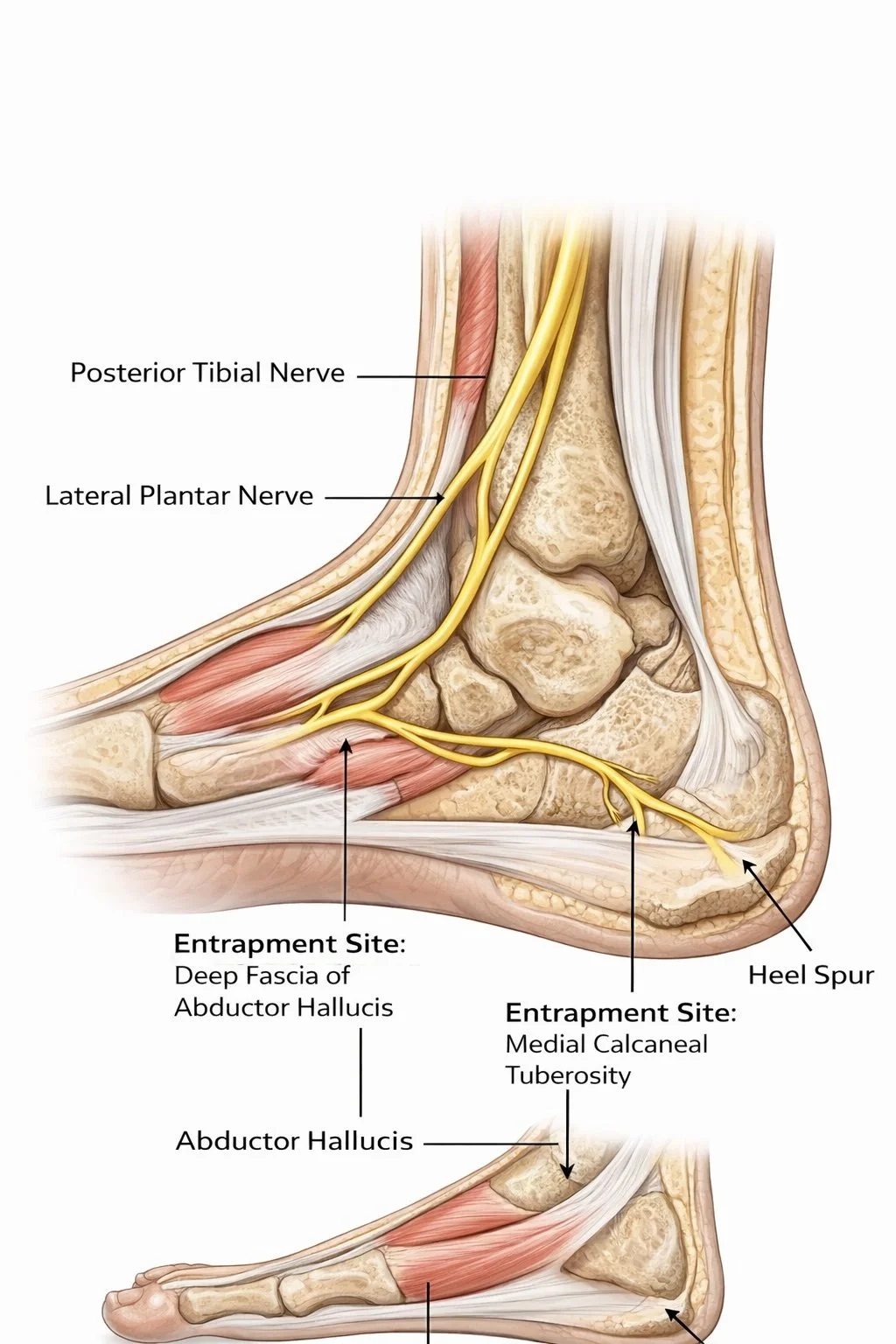
The Unsung Hero: Meet Your Plantar Fascia
Your plantar fascia isn’t just connective tissue; it’s a living cable of collagen, designed to store and release energy. Every time you take a step, it stretches under load (like a bowstring) and then recoils to propel you forward.
It ties together three critical points:
Calcaneus (heel bone)
Metatarsal heads (ball of the foot)
Proximal phalanges (base of the toes)
In this setup, the fascia acts as the rope in our imaginary pulley system, tightening and relaxing in rhythm with each step.
When it’s working well, you move effortlessly.
When it fails—welcome to plantar fasciitis, heel pain, or flat-foot fatigue.
The Physics Behind the Foot: What Is the Windlass Mechanism?
First described by J.H. Hicks (1954), the Windlass Mechanism explains how the plantar fascia tightens as your toes dorsiflex (lift upwards) during the toe-off phase of walking.
As the toes lift:
The fascia winds around the metatarsophalangeal joints (MTPJs)
The calcaneus is drawn closer to the metatarsal heads
The medial longitudinal arch rises
The foot stiffens into a powerful lever
In short, the foot changes gear—from a flexible shock absorber to a rigid springboard.
Three Modes of Windlass: Passive, Reverse, and Active
Most textbooks stop at “the plantar fascia tightens.” But there’s more nuance. The Windlass works in three distinct modes—each essential for healthy gait.
a. Passive Windlass
This is the original Hicks model:
When the big toe dorsiflexes, the fascia tightens without muscular help. The arch lifts naturally, prepping the foot for propulsion. Hicks even demonstrated this in cadaver feet, proving it’s a mechanical (not muscular) response.
b. Reverse Windlass
During early stance, the opposite occurs. As the arch flattens, the plantar fascia elongates and stores elastic energy—like stretching a spring. This stored tension is later released at push-off. It’s the body’s version of regenerative braking.
c. Active Windlass
Muscles join the party.
The flexor hallucis longus (FHL), flexor hallucis brevis (FHB), and intrinsics support the fascia and fine-tune arch tension. In people with weak or over-stretched fascia, the active system compensates—especially in chronic plantar fasciitis or hypermobile feet.
Anatomy of a Built-in Pulley System
Think of your foot as a triangle, with the arch as its peak.
At the base are two pulleys:
Pulley 1: Head of the first metatarsal
Pulley 2: Groove in the heel bone, the sustentaculum tali
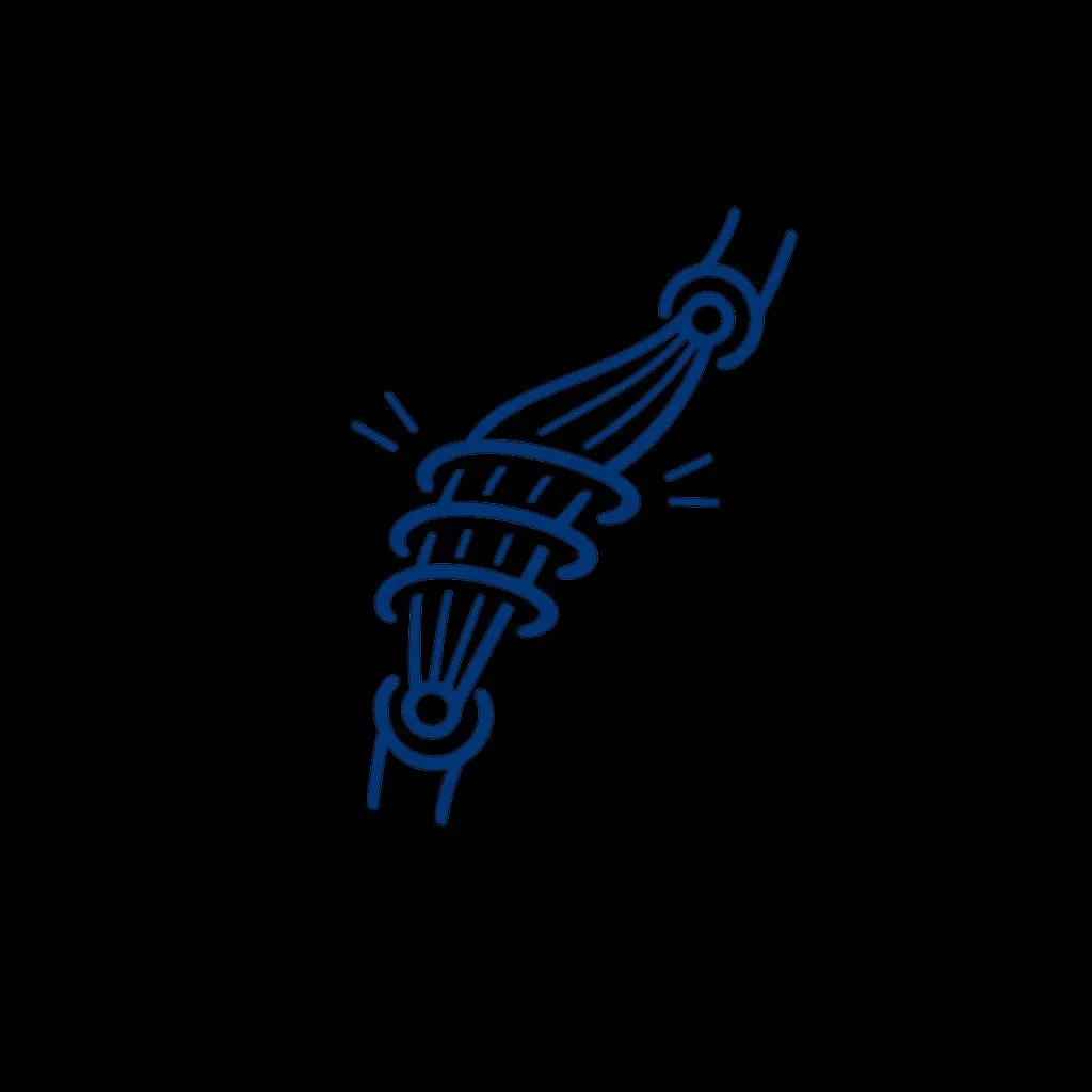
The “rope” running between them is the FHL tendon.
When the big toe lifts, tension pulls the pulleys closer, narrowing the base and forcing the peak (your arch) higher. Voilà—automatic arch lift.
Biomechanically, it’s poetry.
Physically, it’s efficiency.
Clinically, it’s everything.
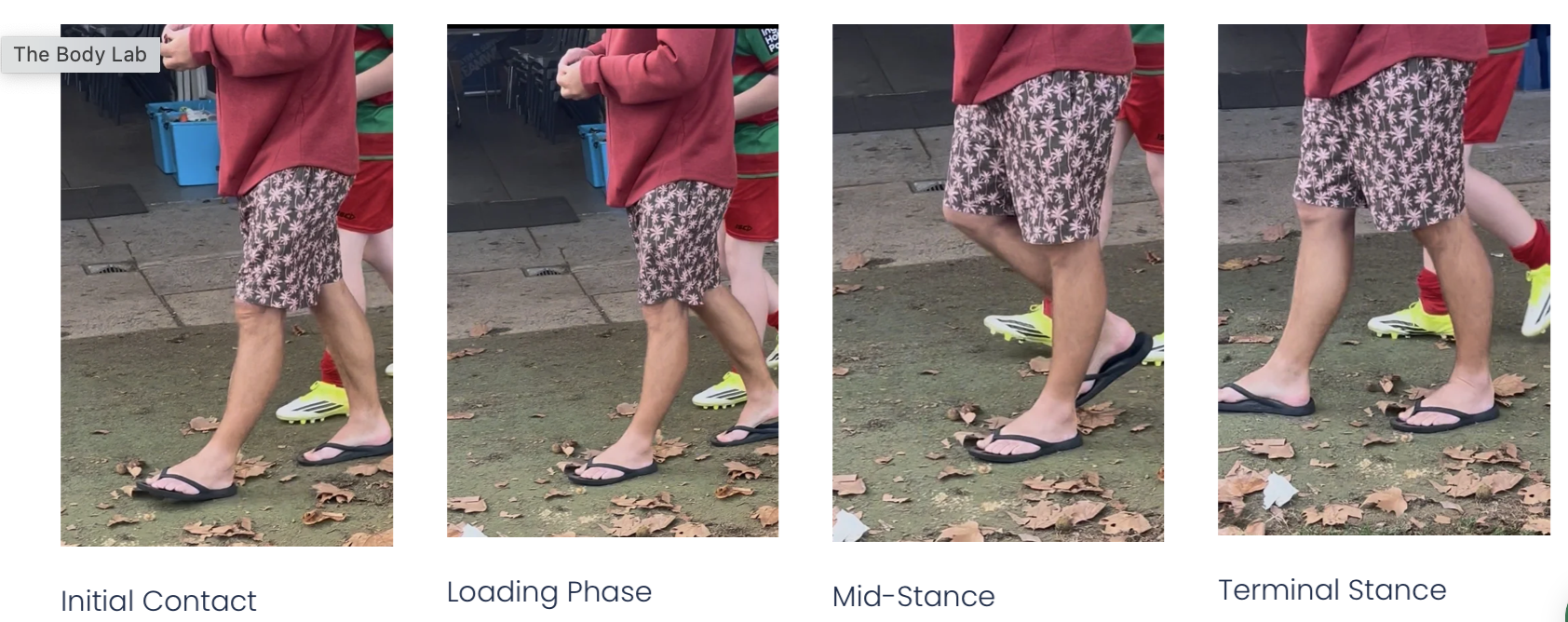
Step by Step: The Windlass in Gait

To make this work perfectly, the big toe needs about 60° of dorsiflexion—with roughly 40° from the toe and 20° from the first metatarsal’s plantarflexion (Neumann, 2013). Too little or too much motion, and efficiency tanks.
When the Windlass Fails: The “Goldilocks Problem”
When your windlass doesn’t engage correctly, your body starts compensating.
Too little toe extension → Foot can’t stiffen → You “pull” your leg forward instead of pushing off.
Too much toe extension → Fascia can’t tension → Same compensation pattern.
Either way, propulsion becomes inefficient, and the chain reaction begins:
Plantar fasciitis (micro-tears at the heel)
Flat-foot pronation and arch collapse
Achilles overload (tendon strain)
Knee valgus or hip over-rotation
Research by Okamura et al. (2020) confirmed that people with heel pain often display reduced windlass function, altered toe dorsiflexion, and delayed arch rise.
Testing the Windlass Mechanism
Clinicians can easily test this:
Have the patient stand and dorsiflex their big toe.
Watch for a visible lift in the arch.
Normal: Arch lifts smoothly (effective fascia tension).
Abnormal: No lift or pain at the medial heel (possible plantar fasciitis).
The Windlass Test (Bolga & Malone, 2004) is a simple but powerful diagnostic tool.
Also check:
Toe range in gait
Midfoot stiffness
Calcaneal motion
Tibialis posterior strength
First MTPJ mobility
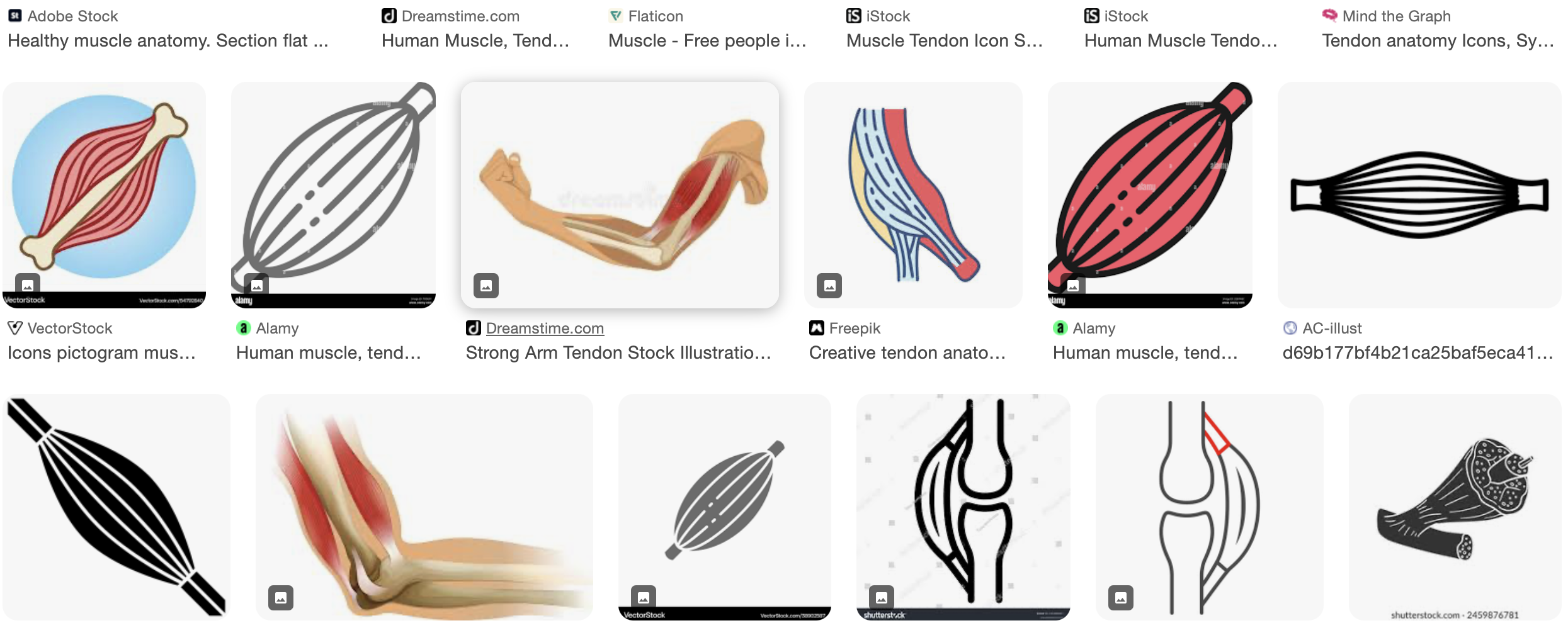
Muscle Allies: The Supporting Cast
The fascia doesn’t work alone.
Key players include:
Flexor Hallucis Longus (FHL): The main “rope.” Wraps around two pulleys and controls toe-off timing.
Flexor Hallucis Brevis (FHB): Stabilises the big toe base.
Tibialis Posterior: Controls pronation and eccentric arch loading.
Peroneus Longus: Anchors the first ray, preventing collapse.
Achilles Tendon: Couples heel rise to fascia strain.
According to Cheng et al. (2008), roughly two-thirds of plantar fascia tension arises from toe dorsiflexion and one-third from Achilles pull—so calf tightness and toe stiffness are equally guilty when the fascia protests.
Clinical Application: Restore the Pulley, Restore the Person
Assess
Observe great toe extension during gait.
Note arch recoil during heel lift.
Identify midfoot stiffness or tibial rotation asymmetry.
Mobilise
Free the first MTPJ to allow dorsiflexion.
Mobilise midfoot and calcaneus to restore glide.
Release tight calf or fascia tissue.
Retrain
Toe Yoga / Short-Foot Drills: Build intrinsic control.
Arch Lift with Knee Rotation: Reactivate fascial recoil.
Heel-Raise with Big Toe Pressure: Re-educate push-off.
When fascia and muscles share the workload, movement becomes efficient, springy, and pain-free.
Why It Matters: Small Toe, Big Impact
When your great toe loses motion or the fascia loses tension, the entire kinetic chain suffers.
Force transfer falters.
Energy leaks upward into the knee, hip, or back.
In their 2022 study, Kappel-Bargas et al. found two distinct windlass patterns:
Immediate Arch Lift: Indicates high fascial stiffness—potential overuse.
Delayed Lift: Suggests under-tensioned fascia—common in flat or hypermobile feet.
Both require targeted mobility and stability work to bring the system back into balance.
Conclusion: The Body’s Quiet Genius
The Windlass Mechanism is nature’s engineering masterpiece—a self-adjusting pulley that lets the foot switch between flexibility and rigidity in milliseconds.
Every step is a symphony of tension and release, arch and toe, fascia and flow.
So next time you’re walking, running, or treating a client with foot pain, remember:
That “stringy bit” in the sole isn’t just connective tissue. It’s a living spring, a mechanical marvel, and a key to effortless motion.
Respect the fascia. Honour the toe. Check the windlass.
Want to Go Deeper?
Explore our upcoming Professional Study Modules at The Body Lab Education, where we unpack fascia dynamics, movement restoration, and 3D foot mechanics with practical, hands-on strategies.
Because understanding the windlass is just the start—mastering it is where transformation begins.
References (Vancouver Style)
Hicks JH. The mechanics of the foot. II. The plantar aponeurosis and the arch. J Anat. 1954;88(1):25–30.
Okamura K et al. Functional biomechanics of the windlass mechanism in symptomatic and asymptomatic individuals. Clin Biomech. 2020;74:13–18.
Caravaggi P et al. The mechanical function of the plantar fascia: a spring in your step. Clin Biomech. 2021;87:7–14.
Kappel-Bargas A et al. Toe dorsiflexion restriction and its relationship to plantar fascial dysfunction: a clinical review. J Foot Ankle Res. 2022;15(1):1–10.
Neumann DA. Kinesiology of the Musculoskeletal System. 3rd ed. Elsevier; 2013.
Cheng HYK et al. Biomechanical analysis of plantar fascia strain in various loading conditions. Foot Ankle Int. 2008;29(9):935–941.
Bolga LA, Malone TR. Plantar fasciitis and the windlass mechanism: A biomechanical link to clinical practice. J Athl Train. 2004;39(1):77–82.
Van Boerum DH, Sangeorzan BJ. Biomechanics of the foot and ankle. Orthop Clin North Am. 2003;34(1):1–17.